
 Submit a Paper
 Propose a Special lssue
 Open Access
ARTICLE
Enhanced Detection of Cerebral Atherosclerosis Using Hybrid Algorithm of Image Segmentation
Department of ECE, RMD Engineering College, Chennai, India
* Corresponding Author: Shakunthala Masi. Email:
Intelligent Automation & Soft Computing 2023, 36(1), 733-744. https://doi.org/10.32604/iasc.2023.025919
Received 08 December 2021; Accepted 02 March 2022; Issue published 29 September 2022
 View Full Text
 Download PDF
Abstract
In medical science for envisaging human body’s phenomenal structure a major part has been driven by image processing techniques. Major objective of this work is to detect of cerebral atherosclerosis for image segmentation application. Detection of some abnormal structures in human body has become a difficult task to complete with some simple images. For expounding and distinguishing neural architecture of human brain in an effective manner, MRI (Magnetic Resonance Imaging) is one of the most suitable and significant technique. Here we work on detection of Cerebral Atherosclerosis from MRI images of patients. Cerebral Atherosclerosis is a cerebral vascular disease causes narrowing of the arteries due to buildup of fatty plaque inside the blood vessels of the brain. It leads to Ischemic stroke if not diagnosed early. Stroke affects majorly old age people and percentage of affected women is more compared to men. Results: Preprocessing is done by using alpha trimmed mean filter which is used to remove noise and also it enhances the image. Segmentation of cerebral atherosclerosis is done by using K-means clustering, Contextual clustering, and proposed Hybrid algorithm. Various parameters like Correlation, Pixel density, energy is determined and from the analysis of parameters it is determined that proposed Hybrid algorithm is efficient.Keywords
Atherosclerosis may cause progressive cerebral ischemia or cerebral vascular insufficiency; it may lead to progression of mental disorder even to stroke, motor impairment, and cognitive disability and even to death [1]. Due to stroke the annual rate of mortality in US is about 800000. Mostly older people are affected by this disease. People with high blood pressure, high cholesterol and smoking habit are more likely to develop atherosclerosis. Signs and symptoms include numbness or weakness of arms or legs, difficult in speech, temporary loss of vision [2]. The most perfused organ in human body is brain, and in human body blood is first supplied to brain, hence the brain tissues are very sensitive to decrease in blood supply. It has been believed over decades that atherosclerosis lesion in major extra and intra cranial branches may cause ischemic disorders. Around 88% of ischemic disorder is due to Atherosclerosis. The progression of cerebrovascular disease is often a sufficiently slow process. Since it spreads widely many arterial branches are affected. Neurodegenerative disorder leads to long chronic ischemia which is slowly progressing that may lead to cognitive impairment, atrophic tissue changes and even mental disorder if treatment for such disorders is not given at early stage of the disease. For an aggravating atherosclerosis lesion a reconstructive transcatheter surgery is required. According to the severity of the disease, treatment process varies. Low risk patients are treated with medications and high-risk patients are recommended for surgical treatment. The poor blood circulation process in blood vessels of human brain leads to the formation of abnormal blood vessels. A mass of semi-solid blood forms the blood clot. In absence of blood clot blood flow is normal and move freely through arteries and veins. Based upon the type of blood clot and its location it is necessary to evaluate the coagulation or blood clot. [1] Majorly two types of brain stroke occur. One is hemorrhage stoke and the other is ischemic stroke. Hypertension, hyperlipidemia, diabetes, micro-emboli caused by carotid arrhythmias plays an important role in its progression.
The quality of image plays a very important role in medical image processing. Based on accuracy of segmentation the quality of medical image diagnosis will be successful. Image quality that provides human body’s unparalleled view is one of the most important factors for identification of atherosclerosis. [3]
In order to detect Cerebral Atherosclerosis, image processing steps like Preprocessing and Segmentation is required. Preprocessing is a technique used to enhance the images and also it removes noise from the image. Different methods like image scaling, Gray scale conversion is used followed by Alpha trimmed mean filter which is used to remove noise. Image normalization techniques like histogram stretching, histogram equalization is performed to increase the contrast of image and Image Sharpening is performed over image to highlight fine details and edges in an image [4]. Image segmentation is used to split the image into meaningful region. It is used to provide distinction between the region we want to inspect further with region in the background. The previous techniques for analyzing brain atherosclerosis are unable to produce better and accurate identification of affected lesion area. The proposed method analyses and segments atherosclerotic lesion area by forming a hybrid algorithm that combines the pros of three major clustering algorithms such as K-means, Contextual Clustering and Particle Swarm Optimization. Proposed method is depicted in flow diagram Fig. 1.

Figure 1: Overall Work Flow diagram of Proposed method
Santichai Fueanggan et al. (2011) [5] proposed a methodology to specify area affected by Ischemic stroke by dividing image data with statistical method and probability into two different areas, they are normal and abnormal areas in the brain. They assign threshold level automatically in CTP, CBV and CBF images. Images are finally divided to non-tissue area, dead tissue area and mismatch area. Finally concluded with brain by separating ischemic stroke area and the results are compared with brain specialist’s suggestion.
Liao et al. (2016) [6] proposed a method for detection and classification of hematoma and type of it using decision rules. Multi resolution Thresholding and region growing methods were used for identification of hematoma region.
Spyretta Golemati et al. (2013) [7] analyzed ultrasound images of carotid a low-cost imaging system. Valuable information such as plaque composition and its stability revealed by Ultrasound images are considered to be the basic information. And further analysis made on atherosclerosis and risk towards Ischemic stroke.
Gharnali et al. (2018) [8] proposed an effective method for segmentation of Magnetic resonance images using FCM technique. FCM an effective object function has been constructed in this technique by replacing Euclidean distance with kernel induced distance. Experiments were performed on synthetic images as well as on MRI images qualitatively and quantitatively using Dice factor for segmentation.
Eero Salli et al. (2001) [9] presented contextual clustering algorithm for calculating SPM (Statistical Parametric maps) from three dimensional image. Neural activations from fMRI images are detected. This divides activation and non-activation area that helps to detect false activation. The system uses a Markov random field prior and Iterated Conditional Modes (ICM) algorithm for the above process.
Chawla et al. (2009) [10] proposed a technique for detection and classification of all types of stroke like acute, chronic and hemorrhagic present in brain images. The abnormalities were detected using two-level classification scheme. Histogram features are used in first level followed by wavelet-based features in second level.
Tang FH (2011) [11] proposed a novel Circular Adaptive Region of Interest (CAROI) method to analyze the Computed Tomography (CT) images of the brain. It is used detect ischemic stroke with small lesions using image feature characteristics in early manner and it is shown that improvement in sensitivity and specificity are achieved. Also, it helps to improve the efficiency and accuracy of clinical practice.
For desired segmentation, a single segmentation method cannot achieve good result usually, this is due to medical image complexity. Adoption of combination of different approach is necessary for better segmentation process [12]. For segmenting the image in a most efficient manner a novel algorithm is formed by combining multiple algorithms together. The Hybrid algorithm includes K-means algorithm, Contextual clustering and Particle Swarm Optimization (PSO) [13].
Partitioning dataset into K predefined subgroups that is non-overlapping is known to be k-means clustering and each data is clustered to any one group. It maintains similarity among inter-cluster data points. Center point should be chosen such that it maintains far distance among the clusters. Data points are assigned to the clusters such that the centroid to data point distance is as minimum as possible. Homogeneity of the data points within a cluster is decided by minimal variation of data points in the cluster.
i) Cluster number specification.
ii) Centroid initialization by sorting dataset followed by K data point selection in a random manner for centroid selection without replacement.
iii) Continue iteration for fixed centroids.
• Squared sum between data points are calculated.
• Data points are assigned to the cluster that is closer to the centroid of the particular cluster.
• Centroid has been computed by averaging every cluster data.
The above approach can also be described as Expectation maximization. Data points are assigned to clusters with respect to the distance between centroids and data points. Followed by computing the real centroid.
A topic of pattern recognition in computer vision is Contextual image segmentation; it is an approach by which images are segmented based on contextual information in it. The approach majorly focuses on relationship of the nearby pixels which is meant as contextual [14]. Segmentation of images with contextual information is the ultimate aim of this approach. A data is segmented into category 1 (ω0) and category 2 (ω1) with Contextual clustering Algorithm. Its assumed that the data is drawn from standard normal distribution. For implementation of contextual clustering the following steps are adopted [15].
i) Definition of Tcc (decision parameter) and β–neighborhood information’s weight. Total number of data in neighborhood is defined by Tn. Total data Zi of ‘I’.
ii) Data classification done with Zi > Tα to category1 ω0 and Zi < Tα to category1 ω1. And classified data of two classes are stored to C0 and C1.
iii) Number of data Dn is counted for each data ‘I’ that belongs to class ω1 in data “I” of neighborhood. Data that are outside the range are assumed belonged to class ω0.
iv) Data classification done with the following equation to ω1 and rest to ω0. Classified data are stored to variable C2.
v. When C2 ≠ C1 and C2 ≠ C0 then data from C1 is copied to C0 and data from C2 is copied to C1. Then to step 3 else return to C2.
• Read image and split to 3X3 windows.
• Pattern is formed by sorting the values of the window.
• Find the patterns median.
• Values greater than Um are identified with count.
• Calculation of CC using
• CC values are finally assigned as the segmented data.
3.3 Particle Swarm Optimization
Particle swarm’s optimization is existing population algorithm. Collection of behavior that is explored by the food’s origin is the basic source of swarm’s technology. Type of fitness function is evaluated for every fitness value that the particle individually also result on site is given by the data in the structure of speed. For majority of area in the given problem the particles are executed in the form of a useful purpose.
The main algorithm begins when special solutions are generated by the compiling molecule that is by scanning using organized iteration gives optimum value. On the basis of higher values repeated modification is done on the particles. Maximum particles like “best” is the term defined for the values found. In the value molecules the second value is determined with the population are called as “gbest”. The arithmetic optimization of existing character is the form of this method. The suggestion is that this configureuration set is applied while in the image segmentation. By using the population research technique, the status of the group is determined. Comparative effortlessness and easiness of use is the main feature of many optimization algorithm. Genetic algorithm refers to many similarities that are shared with scalable computing method. With a set of arbitrary solution, the structure is in progress and by modifying generations the search is improved.
i) Population, speed and location Initialization.
ii) Individual particles best(best) value is evaluated.
iii) Finest fitness value is replaced by improved fitness (pbest) sets the recent value as new Pbest.
iv) The particles that are having excellent fitness value of all the particles as the gbest is chosen.
v) Particle velocity estimated for every particle.
vi) Modification of position of the particle
vii) Stop
Vid = Particle Velocity in d-dimension
Vmin and Vmax Minimum and Maximum distance value
PID = particle position
Pidbest = Individual best
Gidbest = Global best
C1, C2 Constants
i) Image dimensions are fixed to a limit that ranges between 0–255. Dimension of the images are in the range of 0–255.
ii) MRI brain is processed within the upper and lower range.
iii) Preprocessing on brain MRI image with gray scale conversion, denoising with alpha trimmed median filter, and applied with morphological operations.
iv) Initial clustering process has been carried out with K-means clustering. The brain MRI image has been splitted to two clusters.
v) Initialization of affected lesion in image is randomly chosen as initial seed point from result of Kmeans.
vi) For segregating lesion region from the other tissues bounded around and the edema regions, the seed point value acts as the median or center value.
vii) Data classification done with the Eq. (1) to ω1 and ω0.
viii) CC Calculation using Eq. (2)
ix) The process is repeated with step V with repetitive iteration.
x) Then followed by Particle Swarm Optimization for optimizing the clusters with affected region. Thus, helps in exact clustering process.
xi) ROI is the atherosclerosis affected region. Rest are defined by NROI. The flow diagram Fig. 2 of Proposed Hybrid method is as follows:

Figure 2: Proposed hybrid method
Data set consists of various MRI images and it is collected from Madras Scans and research gate and are preprocessed using alpha trimmed mean filter. Input image of patient MSA9 is shown in Fig. 3a, Resized image is shown in Fig. 3b, Denoised image is shown in Fig. 3c, Skull stripped image is shown in Fig. 3d, Morphological opening image is shown in Fig. 3e, Segmentation with multiple iteration image is shown in Fig. 3f, Region of interest result after PSO is shown in Fig. 3g and Affected region is shown in Fig. 3h.

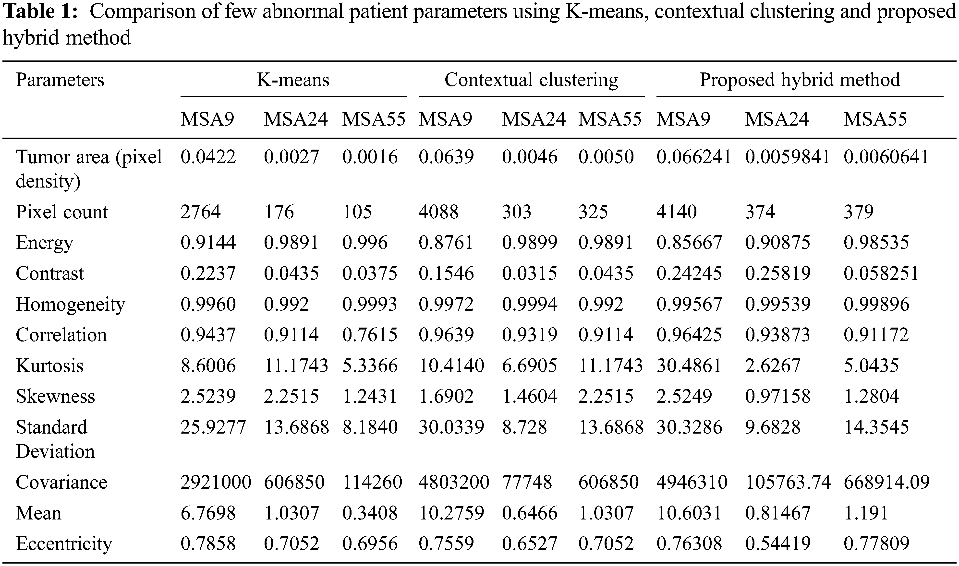
Figure 3: a. Input image, b. Resized image, c. Image after denoising, d. Skull stripped image, e. Morphological opening image, f. Segmentation with multiple iteration, g. Region of interest result of PSO, h. Affected region K-means Clustering, Contextual clustering and Proposed Hybrid method have been applied across input MRI normal and abnormal images and result of patients MSN33, MSA9, MSA24, MSA55 is shown in Fig. 4 and nearly 12 parameters are determined and it is shown in Tabs. 1 and 2. From the Fig. 4 it is observed that proposed hybrid algorithm gives better results as compared to K-means and Contextual clustering algorithm
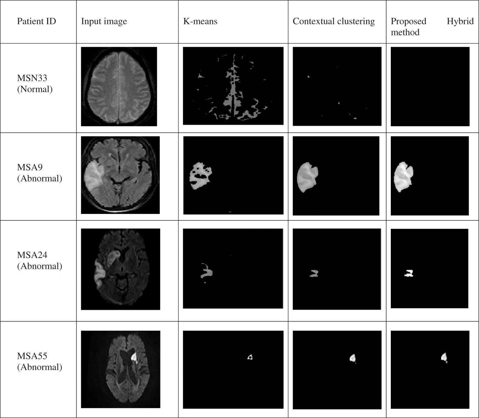
Figure 4: Comparison of different segmentation techniques

The parameters like Tumor area, Pixel count and Correlation of Abnormal patient is taken into account and comparison chart of these parameters using K-Means, Contextual Clustering and Proposed Hybrid method is shown in Figs. 5a–5c and from the chart we could able to analyze tumor area, pixel count and correlation parameter is good in proposed hybrid method.
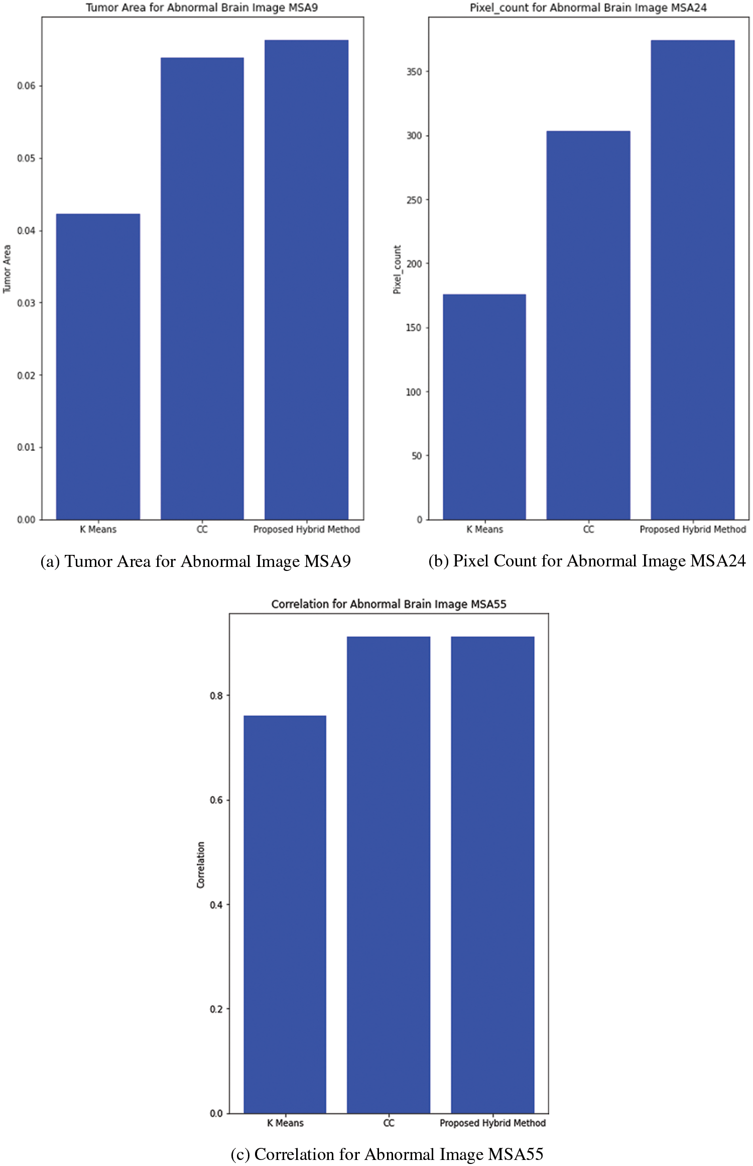
Figure 5: (a) Tumor area for abnormal image MSA9, (b) Pixel count for abnormal image MSA24, (c) Correlation for abnormal image MSA55
The parameters like Tumor area, Pixel count and Correlation of Normal Patient is taken into account and comparison chart of these parameters using K-Means, Contextual Clustering and Proposed Hybrid method is shown in Figs. 6a–6c and from the chart we could able to analyze tumor area, pixel count is zero for proposed method and non-zero for other two methods also correlation parameter is high in proposed hybrid method.
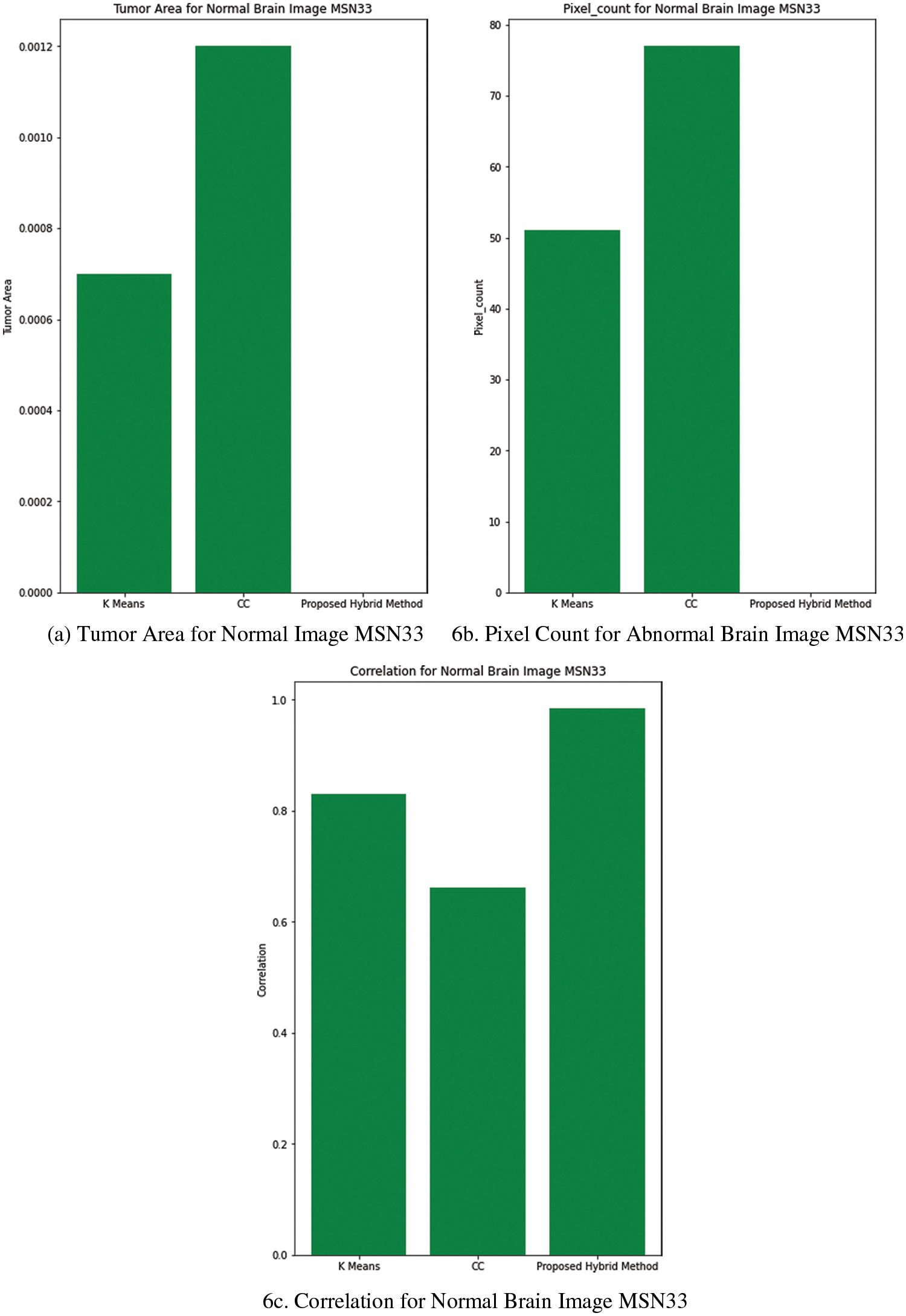
Figure 6: (a) Tumor area for normal image MSN33, (b) Pixel count for abnormal brain image MSN33, (c) Correlation for normal brain image MSN33
For undertaking the brain surgery, detection of affected region plays a major role in brain images. Different algorithms for atherosclerosis lesion removal have been investigated and its detection through various segmentation algorithms is carried out. The pinpoint of the exact location of the lesion can be provided by MRI scan. Based on steps followed on each algorithm the different outputs are obtained. The lesion recognized is evaluated by a performance measures like tumor area, pixel count and correlation. Segmentation of affected brain area with Hybrid combination of K-Means, Contextual clustering and PSO has been performed with successful results in various abnormal MRI images. This novel hybrid algorithm gives better result when compared to existing algorithms like K-means and contextual clustering method in detection of the atherosclerotic lesion.
Funding Statement: The authors received no specific funding for this study.
Conflicts of Interest: The authors declare that they have no conflicts of interest to report regarding the present study.
References
1. T. Saam, T. S. Hatsukami, N. Takaya, B. Chu, H. Underhill et al., “The vulnerable, or high-risk, atherosclerotic plaque: Noninvasive MR imaging for characterization and assessment,” Radiology, vol. 244, no. 1, pp. 64–77, 2007. [Google Scholar]
2. V. Sree and M. Naresh, “Novel Approaches for predicting risk factors of atherosclerosis,” IEEE Journal of Biomedical and Health Informatics, vol. 17, no. 1, pp. 183–189, 2013. [Google Scholar]
3. A. Ajam, “A review on segmentation and modeling of cerebral vasculature for surgical planning,” IEEE Access, vol. 5, pp. 15222–15240, 2017. [Google Scholar]
4. M. Shakunthala and K. HelenPrabha, “Preprocessing analysis of Brain images with Atherosclerosis,” in IEEE International Conference on Electrical, Computer and Communication Technologies (ICECCT), Coimbatore, India, pp. 1–5, 2019. [Google Scholar]
5. S. Fueanggan, S. Chokchaitam and S. Muengtaweepongsa, “Automatic detection of ischemic stroke area from CT perfusion maps cerebral blood volume and cerebral blood flow,” in 2011 International Symposium on Intelligent Signal Processing and Communications Systems (ISPACS), pp. 1–6, 2011. [Google Scholar]
6. C. C. Liao, F. Xiao, J. M. Wong and I. J. Chiang, “A knowledge discovery approach to diagnosing intracranial hematomas on brain CT: Recognition, measurement and classification,,” In: Zhang, D. (eds) Medical Biometrics. ICMB 2008. Lecture Notes in Computer Science, vol. 4901. Berlin, Heidelberg: Springer, pp. 73–82, 2008. [Google Scholar]
7. S. Golemati, A. Gastounioti and K. Nikita, “Toward novel noninvasive and low-cost markers for predicting strokes in asymptomatic carotid atherosclerosis: The role of ultrasound image analysis,” IEEE Transactions on Biomedical Engineering, vol. 60, no. 3, pp. 652–658, 2013. [Google Scholar]
8. B. Gharnali and S. Alipour, “MRI image segmentation using conditional spatial FCM based on kernel-induced distance measure,” Engineering, Technology & Applied Science Research, vol. 8, no. 3, pp. 2985, 2018. [Google Scholar]
9. E. Salli, H. J. Aronen, S. Savolainen, A. Korvenoja and A. Visa, “Contextual clustering for analysis of functional MRI data,” IEEE Transactions on Medical Imaging, vol. 20, no. 5, pp. 403–414, 2001. [Google Scholar]
10. M. Chawla, S. Sharma, J. Sivaswamy and L. Kishore, “A method for automatic detection and classification of stroke from brain CT images,” IEEE Engineering in Medicine and Biology Society, vol. 15, pp. 3581–3584, 2009. [Google Scholar]
11. F. H. Tang, D. K. Ng and D. H. Chow, “An image feature approach for computer-aided detection of ischemic stroke,” Computers in Biology and Medicine, vol. 41, no. 7, pp. 529–536, 2011. [Google Scholar]
12. R. Kaur and G. Singh, “Hybrid technique using PSO and region growing algorithm for brain tumor detection,” in Second International Conference on Inventive Communication and Computational Technologies (ICICCT), pp. 1286–1289, 2018. [Google Scholar]
13. P. Libby, P. M. Ridker and A. Maseri, “Inflammation and atherosclerosis,” Circulation, vol. 105, no. 9, pp. 1135–1143, 2002. [Google Scholar]
14. R. Ross, “The pathogenesis of atherosclerosis—An update,” New England Journal of Medicine, vol. 314, no. 8, pp. 488–500, 2002. [Google Scholar]
15. L. S. Athanasiou, P. S. Karvelis, V. D. Tsakanikas, K. A. Stefanou, K. K. Naka et al., “Pathogenesis of atherosclerosis,” Journal of the American College of Cardiology, vol. 47, no. 8, pp. 7–12, 2006. [Google Scholar]
Cite This Article
 Copyright © 2023 The Author(s). Published by Tech Science Press.
This work is licensed under a Creative Commons Attribution 4.0 International License , which permits unrestricted use, distribution, and reproduction in any medium, provided the original work is properly cited.
 Downloads
 Citation Tools