 Open Access
ARTICLE
Intelligent Firefly Algorithm Deep Transfer Learning Based COVID-19 Monitoring System
1 Information Technology Department, Faculty of Computing and Information Technology, King Abdulaziz University, Jeddah 21589, Saudi Arabia
2 Centre for Artificial Intelligence in Precision Medicines, King Abdulaziz University, Jeddah 21589, Saudi Arabia
3 Mathematics Department, Faculty of Science, Al-Azhar University, Naser City 11884, Cairo, Egypt
4 Department of Medical Microbiology and Parasitology, Faculty of Medicine, King Abdulaziz University, Jeddah 21589, Saudi Arabia
5 Health Promotion Center, King Abdulaziz University, Jeddah 21589, Saudi Arabia
6 Computer and Information Technology Department, The Applied College, King Abdulaziz University, Jeddah 21589, Saudi Arabia
7 Basic Science Department, College of Science and Health Professions, King Saud Bin Abdulaziz University for Health Sciences, Jeddah 21423, Saudi Arabia
8 King Abdullah International Medical Research Center, Ministry of National Guard-Health Affairs, Jeddah 21423, Saudi Arabia
* Corresponding Author: Mahmoud Ragab. Email:
Computers, Materials & Continua 2023, 74(2), 2889-2903. https://doi.org/10.32604/cmc.2023.032192
Received 10 May 2022; Accepted 10 June 2022; Issue published 31 October 2022
Abstract
With the increasing and rapid growth rate of COVID-19 cases, the healthcare scheme of several developed countries have reached the point of collapse. An important and critical steps in fighting against COVID-19 is powerful screening of diseased patients, in such a way that positive patient can be treated and isolated. A chest radiology image-based diagnosis scheme might have several benefits over traditional approach. The accomplishment of artificial intelligence (AI) based techniques in automated diagnoses in the healthcare sector and rapid increase in COVID-19 cases have demanded the requirement of AI based automated diagnosis and recognition systems. This study develops an Intelligent Firefly Algorithm Deep Transfer Learning Based COVID-19 Monitoring System (IFFA-DTLMS). The proposed IFFA-DTLMS model majorly aims at identifying and categorizing the occurrence of COVID19 on chest radiographs. To attain this, the presented IFFA-DTLMS model primarily applies densely connected networks (DenseNet121) model to generate a collection of feature vectors. In addition, the firefly algorithm (FFA) is applied for the hyper parameter optimization of DenseNet121 model. Moreover, autoencoder-long short term memory (AE-LSTM) model is exploited for the classification and identification of COVID19. For ensuring the enhanced performance of the IFFA-DTLMS model, a wide-ranging experiments were performed and the results are reviewed under distinctive aspects. The experimental value reports the betterment of IFFA-DTLMS model over recent approaches.Keywords
Nowadays, the COVID-19 outbreak is considered as a universal problem confronted by healthcare administrations. From November 19, 2020, the overall quantity of individuals globally who confirmed that they were affected with SARS-COV-2 was above 56.4 million, whereas the total quantity of mortality from the COVID-19 is exceeding 1.35 million, so showing that coronavirus infections were increasing globally [1]. The patients who are affected by COVID-19 have numerous symptoms, like decrease in oxygen saturation level, fever, body pain, diarrhea, dry cough, shortness of breath, loss of taste and smell, vomiting, and abnormal pulse rate, headache, and sore throat [2]. Amid such indications, low oxygen saturation level, abnormal pulse rate, and high fever were concerned as serious. Shortness of breath and low oxygen saturation level leads to hypoxia and hypoxemia. Patients suffer from hypoxemia and difficulties with heartbeat are of less chance of living [3]. In some cases, patients will not identify hypoxia and a rising pulse rate and become disease by not having appropriate medication. Non-invasive healthcare can be the only possible means to lower the spreading of virus effectively. Technologies were quickly advancing currently and non-invasive healthcare has grabbed the interest of several researchers worldwide alleviating the work pressure on health labors [4]. Also, the progression of healthcare technologies might substantially diminish the amount of useful clinical sources.
An automatic health monitoring module was enhanced which creates a warning in the serious situation of the patient [5]. A chest radiology image-oriented detection technique could prevail numerous benefits rather than classical techniques. It is quick, to examine many cases concurrently, has better accessibility, and most significantly this technique is more helpful in clinics not having few or restricted testing kits and sources [6,7]. Also, providing significance of radiography in latest healthcare scheme, radiology imaging system was existing in all clinics, thus makes radiography-related techniques easily available and very convenient. Authors are aiming at deep learning (DL) methods for identifying certain particular features from chest radiography images of COVID19 cases [8]. Recently, DL made successful accomplishments in several visual tasks that also involves medical image examination. DL has transformed automated disease prognosis and administration by precisely identifying examining, and categorizing paradigms in healthcare images [9,10]. The motive behind this accomplishment is DL methods do not depend on automatic hand-engineered features but such methods study features mechanically through information itself.
Hassantabar et al. [11] employed 3 DL-related techniques for the identification and detection of coronavirus patient using X-Ray images of lungs. For the disease diagnosis, there are 2 methods that are CNN and DNN on the fractal feature of images methodologies by using lung images, straightforwardly. Rasheed et al. [12] inspected the efficiency of ML methodologies for automated prognosis of coronavirus with higher accurateness from X-ray images. Two frequently utilized classifiers have been chosen one is CNN and another one is logistic regression (LR). The first and foremost reason is to make the scheme faster and more effective. Additionally, a dimension reduction method has been inspected grounded on PCA to additionally fasten the process of learning and improve the categorization accuracy through choosing the extremely discriminative feature.
In [13], an original DL-related system, known as COVID-XNet, was exhibited for coronavirus prognosis in chest X-ray images. The presented technique executes a set of preprocessing methods to the input image for variability reduction and contrast enhancement that are fed to a custom CNN for extracting appropriate features and accomplishing the categorization among normal cases and coronavirus. In [14], a novel method for automatic COVID19 recognition with the help of raw chest X-ray images was submitted. The presented method has been enhanced to offer precise diagnostics for multi-class and binary categorization. The DarkNet method can be utilized in this article as a classification for you-only-look-once (YOLO) real time object recognition module. Yoo et al. [15] examine the possibility of utilizing a DL-related decision-tree classifier for recognizing COVID19 from CXR images. The suggested classifier has 3 binary DTs, all of these are trained by a DL method with CNN related method on the PyTorch frame. The primary DT categorizes the CXR images into abnormal or normal. Almalki et al. [16] recommend an original methodology CoVIRNet (COVID Inception-ResNet model), that uses the chest X-rays for diagnosing the coronavirus patient mechanically. The suggested method contains distinct inception residual blocks which cater to information with the help of distinct depth feature mappings at diverse scales, with several layers. The features were concatenated at every suggested classification block, with the help of the average pooling layer, and concatenated feature was passed to the completely linked layer.
This study develops an Intelligent Firefly Algorithm Deep Transfer Learning Based COVID-19 Monitoring System (IFFA-DTLMS). The proposed IFFA-DTLMS model primarily applies densely connected networks (DenseNet121) model to produce a collection of feature vectors. In addition, the firefly algorithm (FFA) is applied for hyper parameter optimization of the DenseNet121 architecture. Moreover, Autoencoder-LSTM (AE-LSTM) model is exploited for the diagnosis and classification of COVID-19. For ensuring the enhanced performance of the IFFA-DTLMS model, a wide range of experiments were performed and the outcomes are reviewed under distinctive aspects.
The rest of the paper is organized as follows. Section 2 offers the proposed model, Section 3 validates the performance, and Section 4 concludes the study.
In this study, a new IFFA-DTLMS model was designed to categorize and identify the occurrence of COVID-19 on chest radiographs. The presented IFFA-DTLMS model primarily applied DenseNet121 model to generate a collection of feature vectors. In addition, the FFA is applied for hyper parameter optimization of the DenseNet121 model. Moreover, AE-LSTM model is exploited for the diagnosis and classification of COVID-19.
2.1 DenseNet Feature Extraction
In this work, the presented IFFA-DTLMS model primarily applied DenseNet121 model to generate a collection of feature vectors. The DenseNet-121 framework is applied as the baseline in this study. Also, transfer learning model takes place in DenseNet module to augment the efficiency of system [17]. In contrast to DenseNet, it needs fewer parameters than that of classical CNN model because they don’t need to learn unwanted feature maps. The fundamental concept of DenseNet structure is the feature reutilization that leads to highly compact form. Subsequently, it requests fewer parameters than that of other CNN models because no feature map is reiterated. When CNN goes further, they face problems. DenseNet makes the connectivity far simpler by easily connecting each layer with other layers. DenseNet utilizes the network ability by reusing features. Fig. 1 depicts the framework of DenseNet. Every single layer in DenseNet obtains input over each preceding layer and transfers feature map to the subsequent layer.

Figure 1: Structure of DenseNet
2.2 Hyperparameter Optimization
Here, the FFA is applied for hyperparameter optimization of the DenseNet121 model. The FFA was initially developed by Yang in [18]. This popular approach has been stimulated by flashing characteristics and social behavior. Because the real-time firefly ecosystem is sophisticated and very complex, the FFA metaheuristic is their calculation. The real formula of attraction is a critical problem that needs to be tackled in the execution of FFA. Generally, the attraction of firefly is depending on the brightness that sequentially depends on the decision criterion. For minimization problem, the brightness of firefly on the x position is evaluated as follows:
In Eq. (1),
In Eq. (2),
Attractiveness
In Eq. (4), the
The subjective firefly i move towards the novel location (in the
where para
In which,
In Eq. (8), D indicates the parameter number. The suitable value for problem is
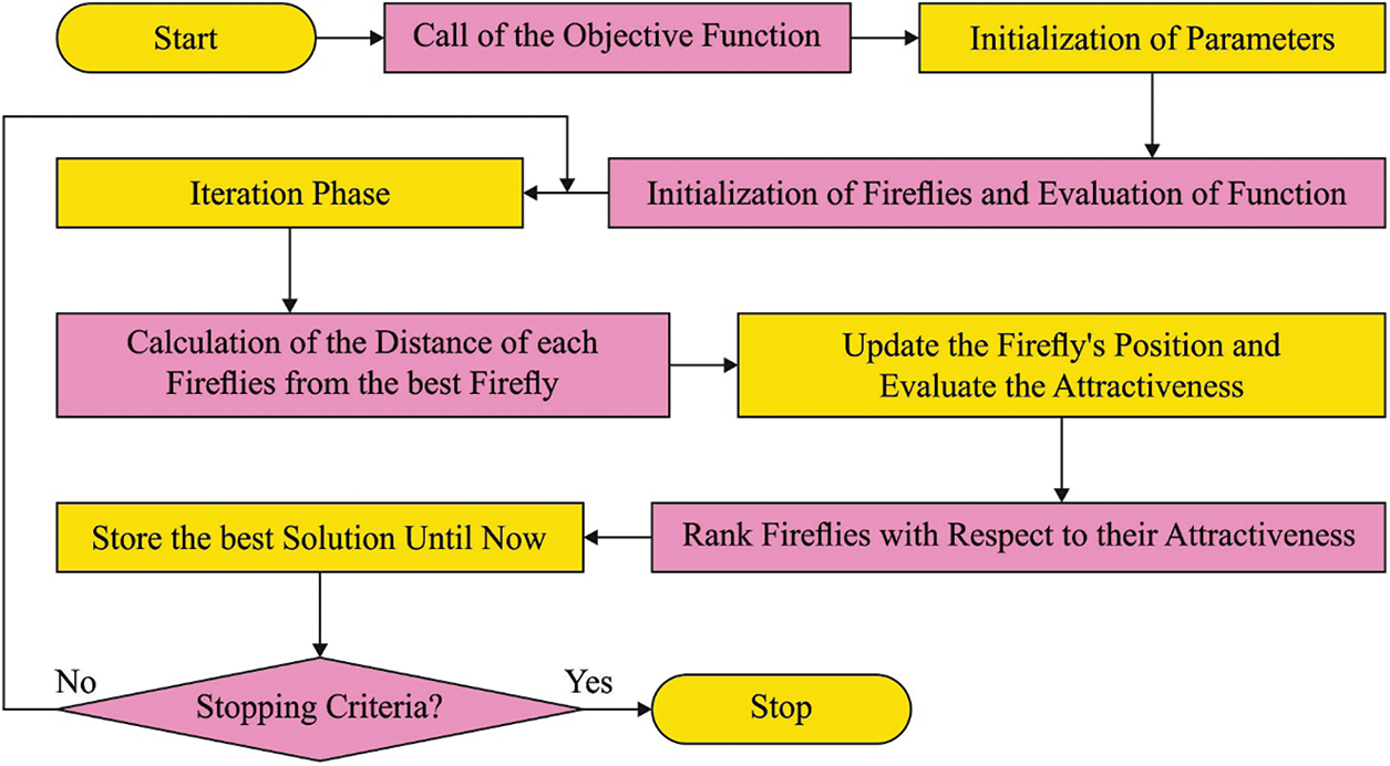
Figure 2: Flowchart of FFA
The FFA approach develops a fitness function (FF) for gaining higher classifier performances. It solves a positive integer for demonstrating the best performance of solution candidate. In such cases, the minimalized classifier error rate is considered as FF in Eq. (9). A worst outcome gains an improved error rate and best possible solution attains a lesser error rate.
2.3 AE-LSTM Based Classification
In the final step, the AE-LSTM model is exploited for the diagnosis and classification of COVID-19. The LSTM model is superior RNN that is initially developed. RNN is a neural network (NN) with recurring relationships among the neurons which allows them to learn from the previous and the current dataset for finding a good outcome [19]. But once the two cells in RNN are farther from one another, it is complex to attain valuable dataset as a result of the gradient explosion and vanishing issues. The solution to this is distinctive neuron termed memory cell. With this neuron, the LSTM is capable of storing effective dataset within a certain duration. Furthermore, LSTM cell is capable of learning what information must be erased, read, and stored from the memory by regulating three dissimilar controlled gates, such as output gate
The AE-LSTM developed comprises LSTM and AE [20]. An AE is an unsupervised neural network in which output and input layers possess similar sizes. AE tries to acquire the individuality function such that the x input is almost analogous to the
Solar power generation predicting
(i) Encoder, wherein the layer is decreased from input to the hidden states and
(ii) Decoder, wherein the neuron in the encoder are imitated.
Consequently, an AE is capable of learning and discovering the connections in the input feature and the distinct architecture of the dataset. The ending outcome of AE-LSTM is an ML technique which is capable of predicting the occurrence of COVID19 by means of input image.
The experimental validation of the IFFA-DTLMS model is tested using the CXR image dataset [21], containing 32224 samples under COVID and 6448 samples under healthy class. A few image samples are illustrated in Fig. 3.
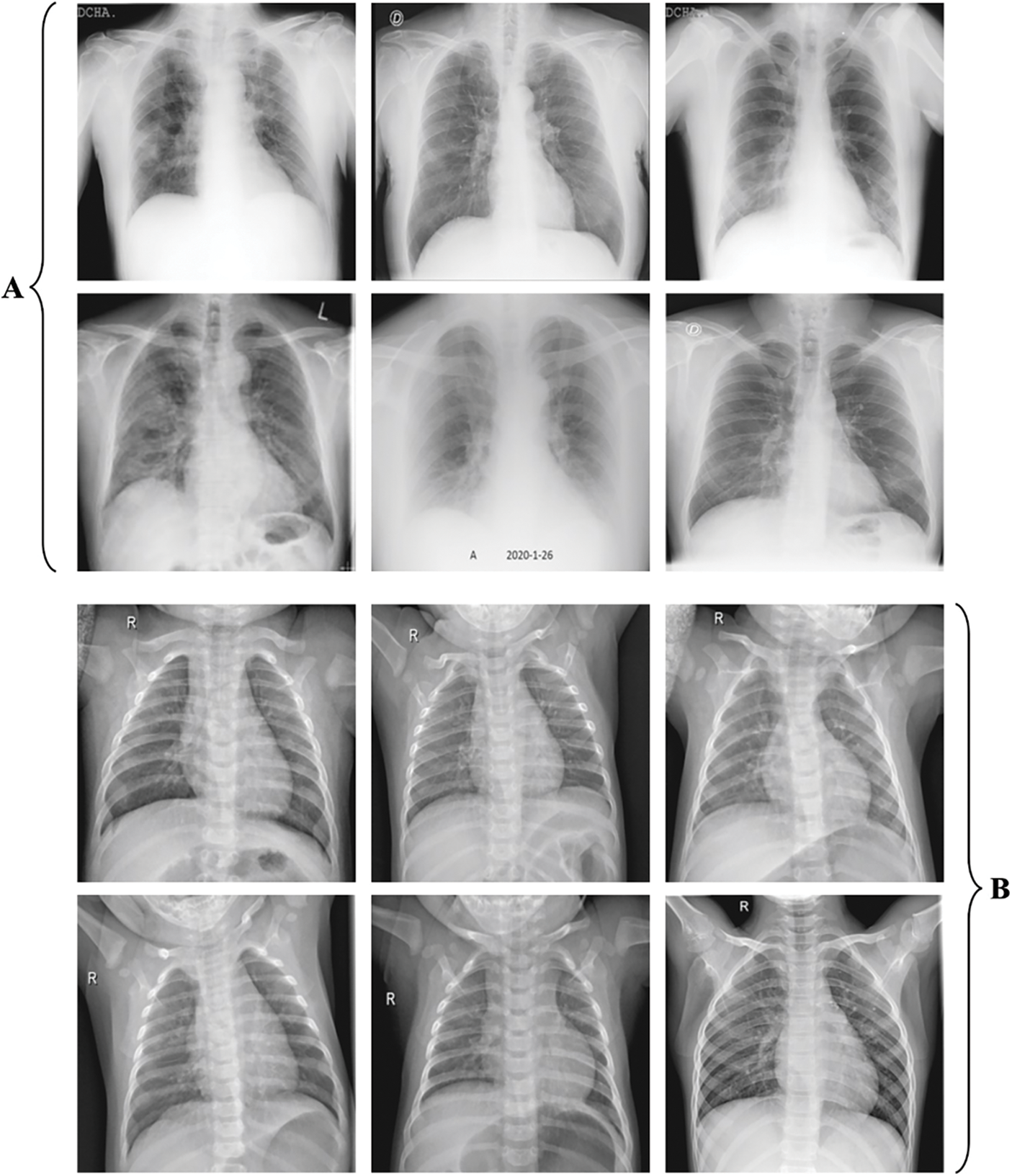
Figure 3: a) COVID b) Healthy
The confusion matrix produced by the IFFA-DTLMS model on the testing dataset is depicted in Fig. 4. On run-1, the IFFA-DTLMS model has recognized 3168 samples into COVID and 3177 samples under healthy. Also, on run-3, the IFFA-DTLMS technique has recognized 3194 samples into COVID and 3146 samples under healthy. Besides, on run-5, the IFFA-DTLMS approach has recognized 3188 samples into COVID and 3153 samples under healthy.
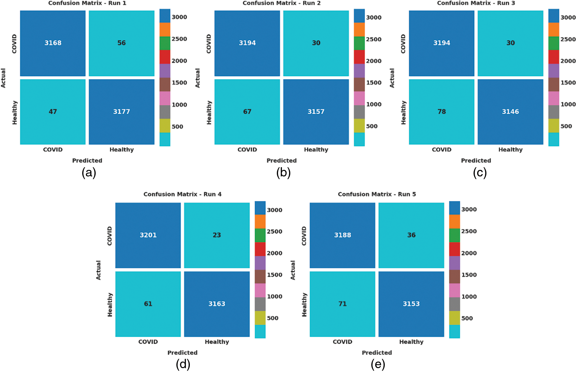
Figure 4: Confusion matrices of IFFA-DTLMS technique (a) Run-1, (b) Run-2, (c) Run-3, (d) Run-4, and (e) Run-5
Tab. 1 and Fig. 5 highlight the overall analysis result of the IFFA-DTLMS model on the testing data. The results portrayed that the IFFA-DTLMS model has reached enhanced performance under every run. For example, on run-1, the IFFA-DTLMS model has provided enhanced average

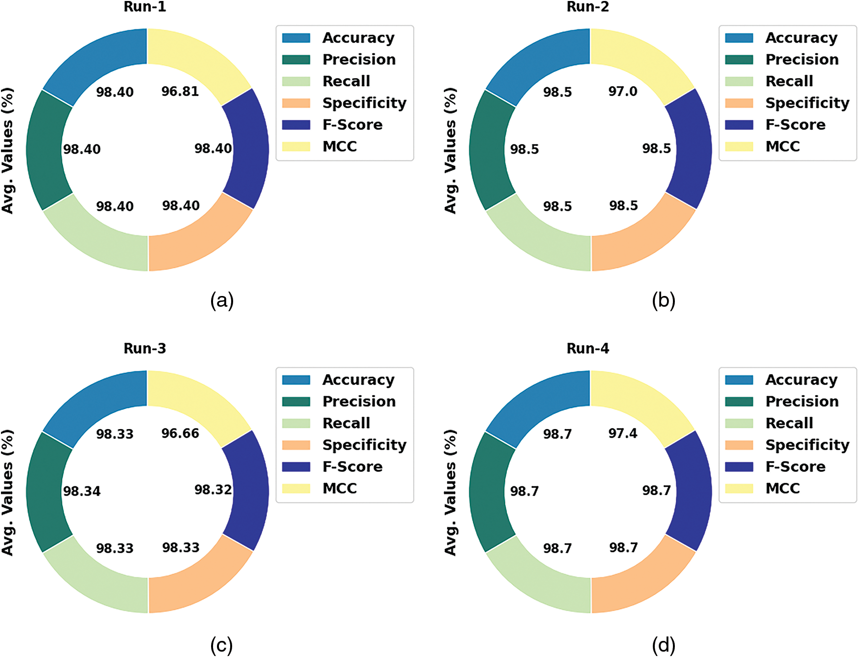
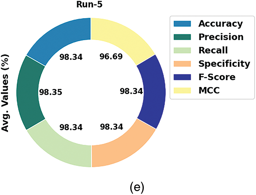
Figure 5: Average analysis of IFFA-DTLMS method (a) Run-1, (b) Run-2, (c) Run-3, (d) Run-4, and (e) Run-5
The training accuracy (TA) and validation accuracy (VA) attained by the IFFA-DTLMS approach on testing data is illustrated in Fig. 6. The experimental outcome implied that the IFFA-DTLMS system has acquired maximum values of TA and VA. In specific, the VA seemed to be higher than TA.
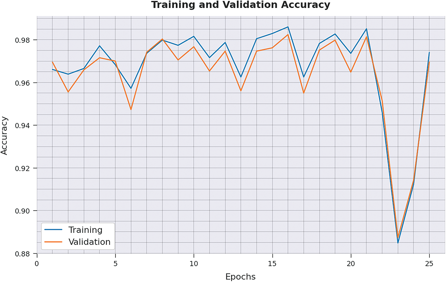
Figure 6: TA and VA analysis of IFFA-DTLMS approach
The training loss (TL) and validation loss (VL) achieved by the IFFA-DTLMS methodology on test dataset are established in Fig. 7. The experimental outcome inferred that the IFFA-DTLMS approach has established least values of TL and VL. In specific, the VL seemed to be lesser than TL.
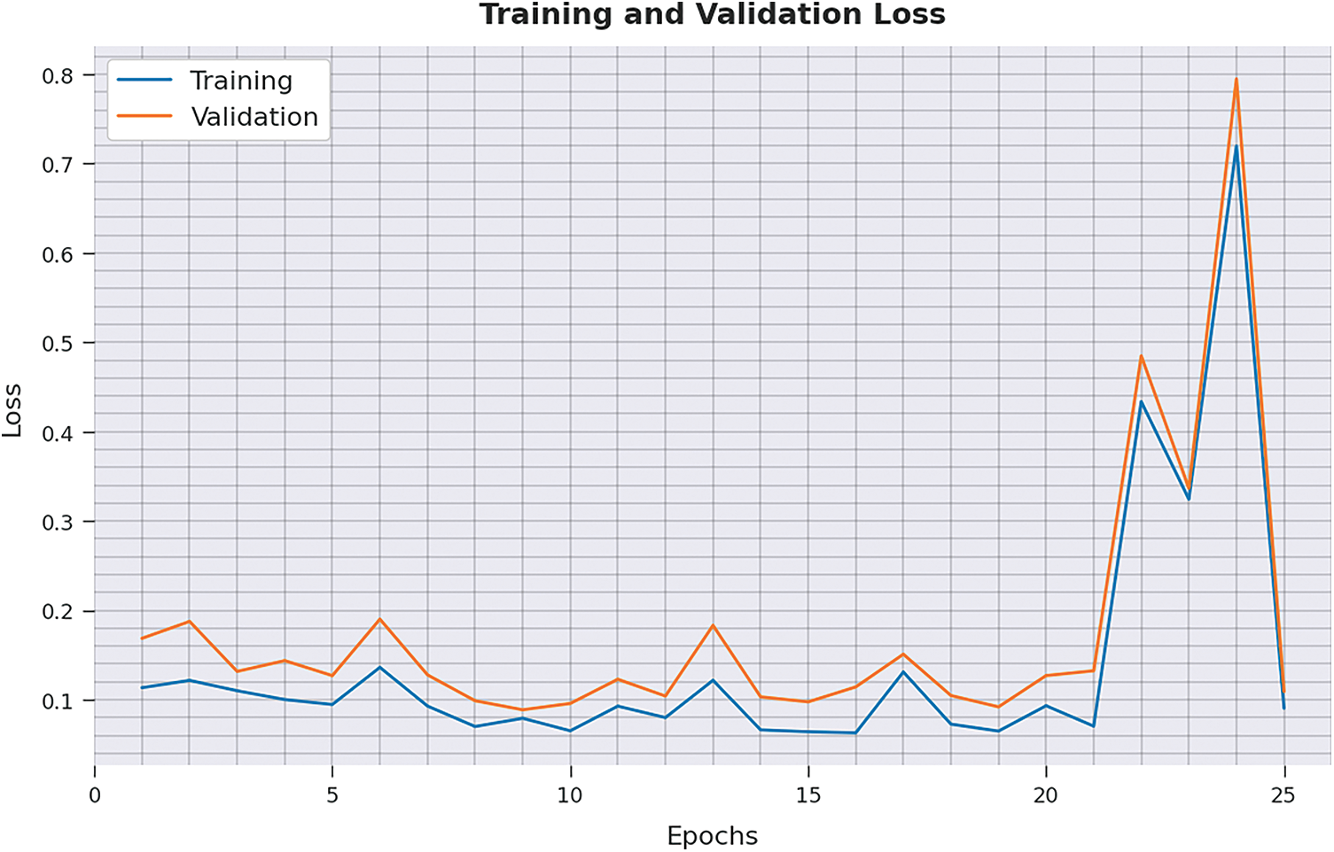
Figure 7: TL and VL analysis of IFFA-DTLMS approach
A brief precision-recall inspection of the IFFA-DTLMS method on test dataset is represented in Fig. 8. By observing the figure, it is noticed that the IFFA-DTLMS model has established maximum precision-recall performance under distinct classes.
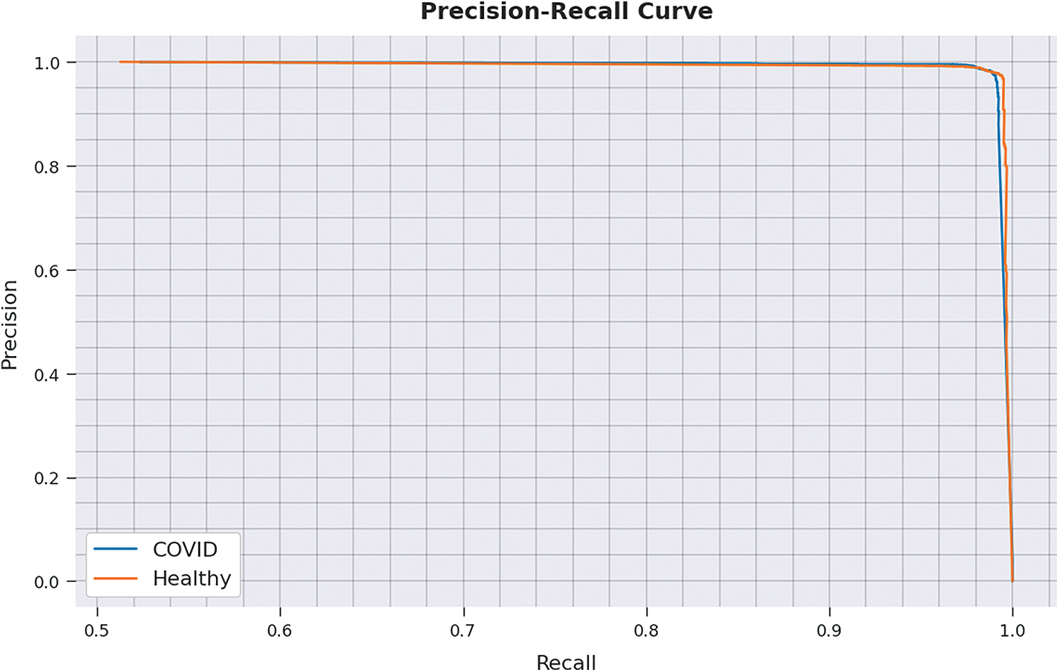
Figure 8: Precision-recall curve analysis of IFFA-DTLMS approach
A detailed ROC investigation of the IFFA-DTLMS methodology on test data is portrayed in Fig. 9. The results specified that the IFFA-DTLMS system has shown its ability in categorizing two distinct classes on test dataset.
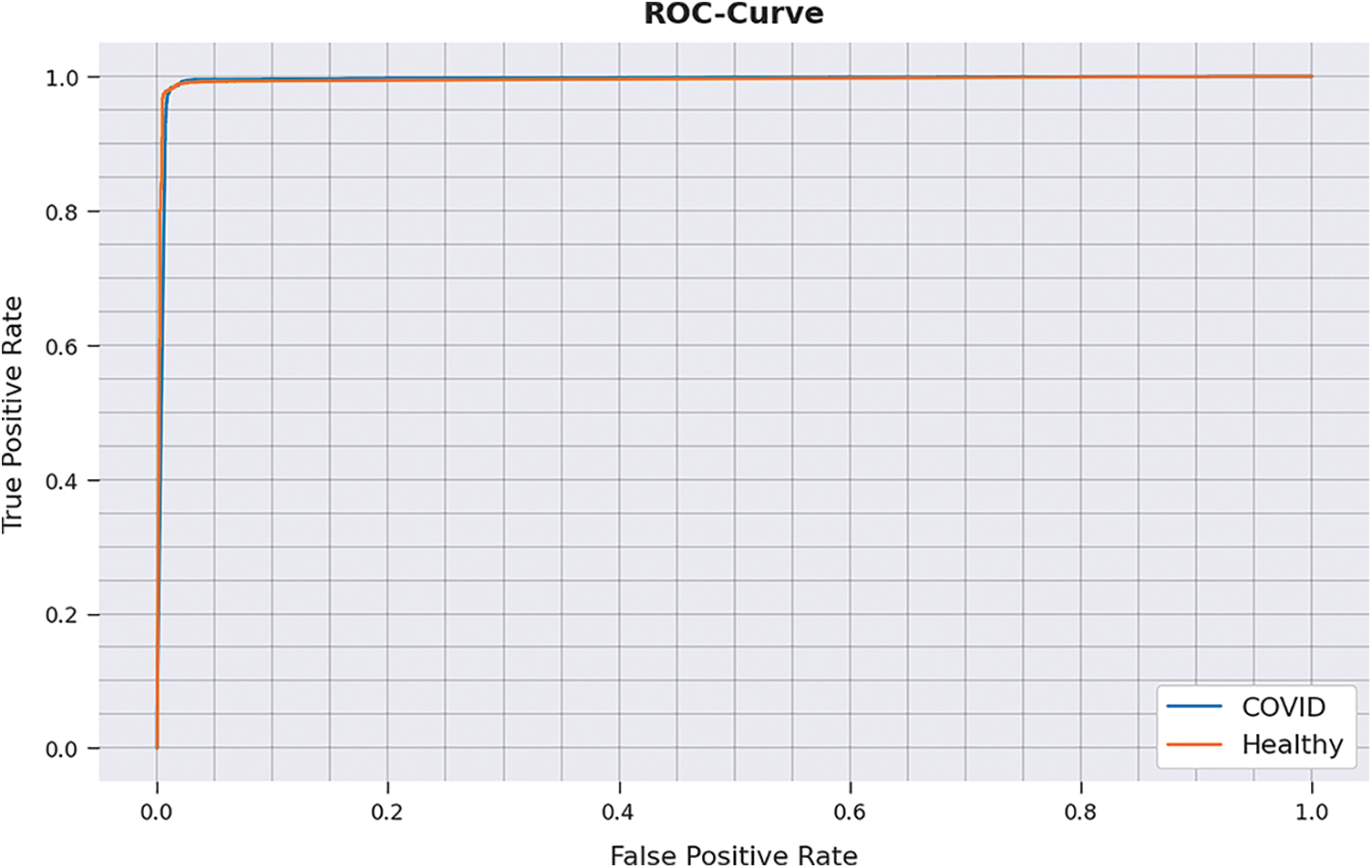
Figure 9: ROC curve analysis of IFFA-DTLMS approach
Tab. 2 illustrates a comparative study of IFFA-DTLMS model with existing models [22–26].

Fig. 10 reports a comparison study of the IFFA-DTLMS model with other models interms of
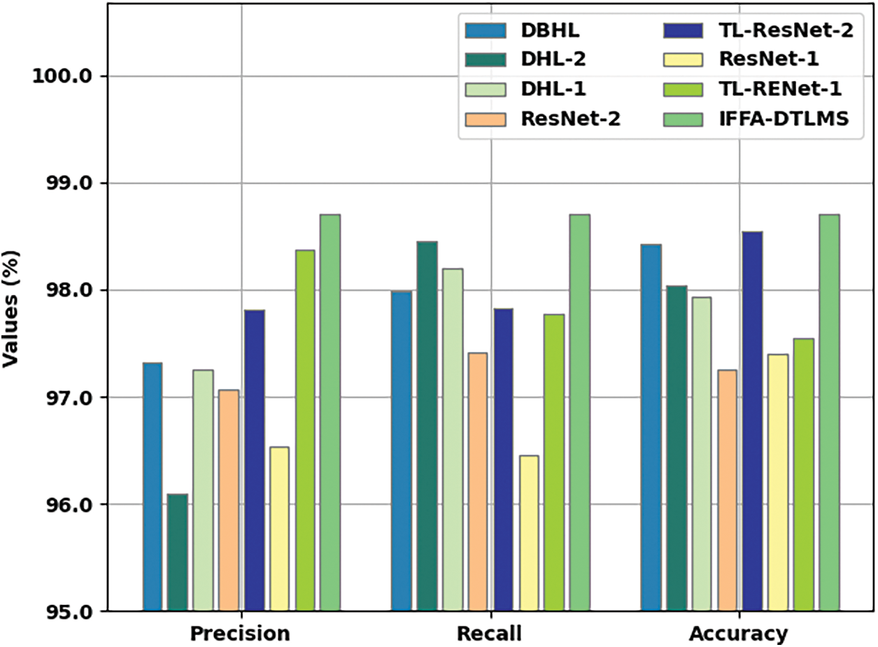
Figure 10:
Fig. 11 reports a comparison study of the IFFA-DTLMS method with other models interms of
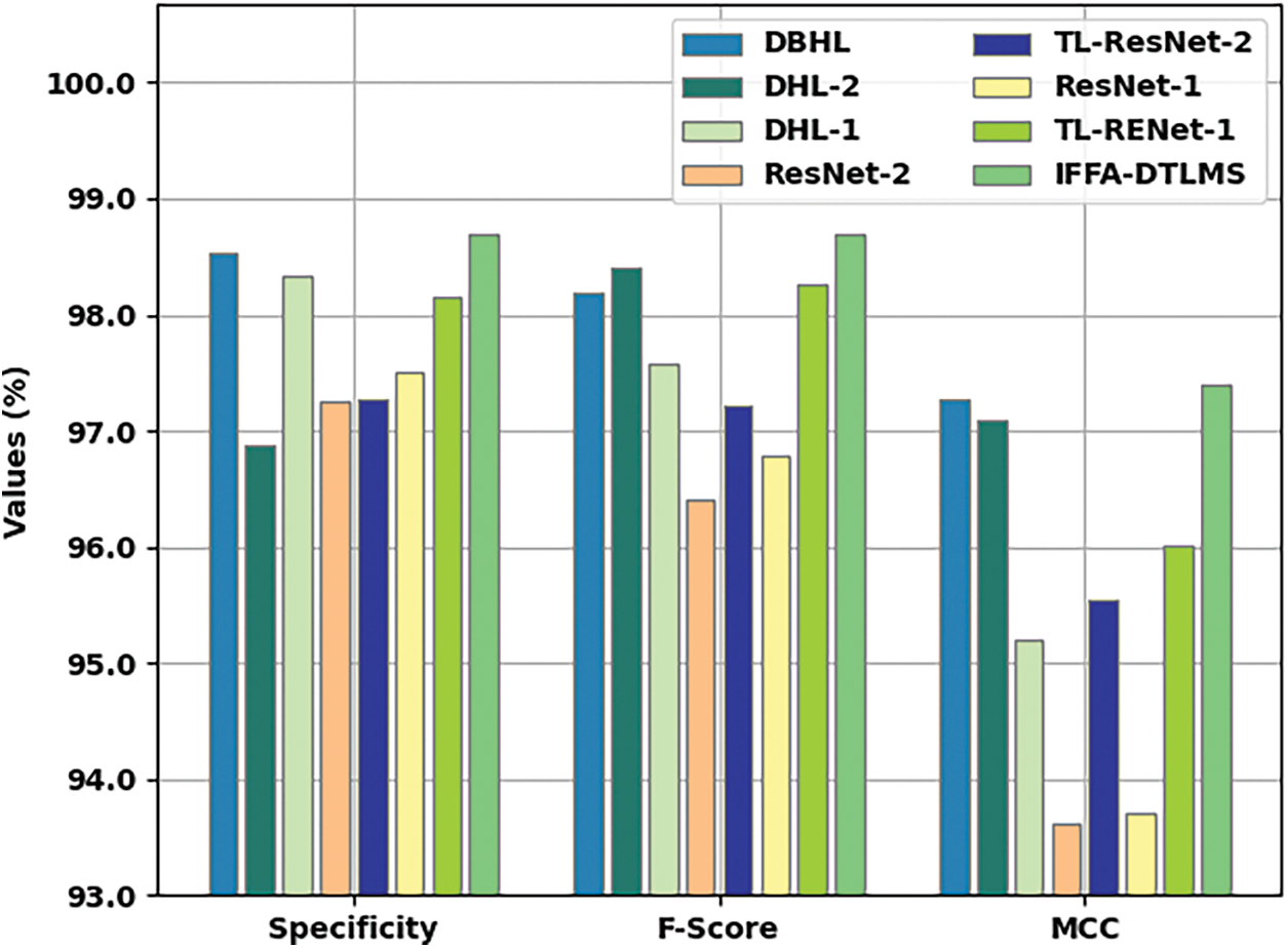
Figure 11:
In this study, a new IFFA-DTLMS model was designed to categorize and identify the occurrence of COVID-19 on chest radiographs. The presented IFFA-DTLMS model primarily applied DenseNet121 model to generate a collection of feature vectors. In addition, the FFA is applied for hyper parameter optimization of the DenseNet121 model. Moreover, AE-LSTM model is exploited for the diagnosis and classification of COVID-19. For ensuring the enhanced performance of the IFFA-DTLMS model, a wide-ranging experiment were performed and the results are inspected under distinct aspects. The experimental value reports the betterment of IFFA-DTLMS model over recent approaches. In the context of future development, the presented IFFA-DLTMS model can be extended to the utilization of computed tomography (CT) images.
Funding Statement: This work was funded by the Deanship of Scientific Research (DSR), King Abdulaziz University, Jeddah, under grant no. (G: 366-140-38). The authors gratefully acknowledge DSR technical and financial support.
Conflicts of Interest: The authors declare that they have no conflicts of interest to report regarding the present study.
References
1. A. Borkowski, “Using artificial intelligence for covid-19 chest x-ray diagnosis,” Federal Practitioner, vol. 37, no. 9, pp. 398–404, 2020. [Google Scholar]
2. R. Yasin and W. Gouda, “Chest X-ray findings monitoring COVID-19 disease course and severity,” Egyptian Journal of Radiology and Nuclear Medicine, vol. 51, no. 1, pp. 193, 2020. [Google Scholar]
3. A. I. Khan, J. L. Shah and M. M. Bhat, “CoroNet: A deep neural network for detection and diagnosis of COVID-19 from chest x-ray images,” Computer Methods and Programs in Biomedicine, vol. 196, no. 2020, pp. 1–9, 2020. [Google Scholar]
4. L. A. Rousan, E. Elobeid, M. Karrar and Y. Khader, “Chest x-ray findings and temporal lung changes in patients with COVID-19 pneumonia,” BMC Pulmonary Medicine, vol. 20, no. 1, pp. 1–17, 2020. [Google Scholar]
5. A. Borghesi and R. Maroldi, “COVID-19 outbreak in Italy: Experimental chest X-ray scoring system for quantifying and monitoring disease progression,” Radiol Med., vol. 125, no. 5, pp. 509–513, 2020, https://doi.org/10.1007/s11547-020-01200-3. [Google Scholar]
6. S. Tabik, A. G. Ríos, J. L. M. Rodríguez, I. S. García, M. R. Area et al., “COVIDGR dataset and covid-sdnet methodology for predicting covid-19 based on chest x-ray images,” IEEE Journal of Biomedical and Health Informatics, vol. 24, no. 12, pp. 3595–3605, 2020. [Google Scholar]
7. E. Benmalek, J. Elmhamdi and A. Jilbab, “Comparing CT scan and chest X-ray imaging for COVID-19 diagnosis,” Biomedical Engineering Advances, vol. 1, no. 2021, pp. 1–6, 2021. [Google Scholar]
8. X. Qi, L. G. Brown, D. J. Foran, J. Nosher and I. Hacihaliloglu, “Chest X-ray image phase features for improved diagnosis of COVID-19 using convolutional neural network,” International Journal of Computer Assisted Radiology and Surgery, vol. 16, no. 2, pp. 197–206, 2021. [Google Scholar]
9. M. Turkoglu, “COVIDetectioNet: COVID-19 diagnosis system based on X-ray images using features selected from pre-learned deep features ensemble,” Applied Intelligence, vol. 51, no. 3, pp. 1213–1226, 2021. [Google Scholar]
10. K. K. Singh, M. Siddhartha and A. Singh, “Diagnosis of coronavirus disease (COVID-19) from chest X-ray images using modified XceptionNet,” Romanian Journal of Information Science and Technology, vol. 23, no. 657, pp. 91–115, 2020. [Google Scholar]
11. S. Hassantabar, M. Ahmadi and A. Sharifi, “Diagnosis and detection of infected tissue of COVID-19 patients based on lung x-ray image using convolutional neural network approaches,” Chaos, Solitons & Fractals, vol. 140, pp. 1–11, 2020. [Google Scholar]
12. J. Rasheed, A. A. Hameed, C. Djeddi, A. Jamil and F. Al-Turjman, “A machine learning-based framework for diagnosis of COVID-19 from chest X-ray images,” Interdisciplinary Sciences: Computational Life Science, vol. 13, no. 1, pp. 103–117, 2021. [Google Scholar]
13. L. D. Lopez, J. P. D. Morales, J. C. Jaime, S. V. Diaz and A. L. Barranco, “COVID-XNet: A custom deep learning system to diagnose and locate covid-19 in chest x-ray images,” Applied Sciences, vol. 10, no. 16, pp. 5683, 2020. [Google Scholar]
14. T. Ozturk, M. Talo, E. A. Yildirim, U. B. Baloglu, O. Yildirim et al., “Automated detection of COVID-19 cases using deep neural networks with X-ray images,” Computers in Biology and Medicine, vol. 121, pp. 103792, 2020. [Google Scholar]
15. S. H. Yoo, H. Geng, T. L. Chiu, S. K. Yu, D. C. Cho et al., “Deep learning-based decision-tree classifier for covid-19 diagnosis from chest x-ray imaging,” Frontiers in Medicine, vol. 7, pp. 427, 2020. [Google Scholar]
16. Y. E. Almalki, A. Qayyum, M. Irfan, N. Haider, A. Glowacz et al., “A novel method for covid-19 diagnosis using artificial intelligence in chest x-ray images,” Healthcare, vol. 9, no. 5, pp. 1–12, 2021. [Google Scholar]
17. T. Chauhan, H. Palivela and S. Tiwari, “Optimization and fine-tuning of DenseNet model for classification of COVID-19 cases in medical imaging,” International Journal of Information Management Data Insights, vol. 1, no. 2, pp. 1–12, 2021. [Google Scholar]
18. X. S. Yang, “Firefly algorithm, stochastic test functions and design optimisation,” International Journal of Bio-Inspired Computation, vol. 2, no. 2, pp. 1–12, 2010. [Google Scholar]
19. J. Zheng, C. Xu, Z. Zhang and X. Li, “Electric load forecasting in smart grids using long-short-term-memory based recurrent neural network,” in 2017 51st Annual Conf. on Information Sciences and Systems (CISS), Baltimore, MD, USA, pp. 1–6, 2017. [Google Scholar]
20. A. Sagheer and M. Kotb, “Unsupervised pre-training of a deep lstm-based stacked autoencoder for multivariate time series forecasting problems,” Scientific Reports, vol. 9, no. 1, pp. 19038, 2019. [Google Scholar]
21. “Dataset: https://github.com/ieee8023/covid-chestxray-dataset. [Google Scholar]
22. M. Ragab, S. Alshehri, N. Alhakamy, W. Alsaggaf, H. Alhadrami et al., “Machine learning with quantum seagull optimization model for covid-19 chest x-ray image classification,” Journal of Healthcare Engineering, vol. 2022, pp. 1–13, 2022. [Google Scholar]
23. R. F. Mansour, J. E. Gutierrez, M. Gamarra, D. Gupta, O. Castillo et al., “Unsupervised deep learning based variational autoencoder model for covid-19 diagnosis and classification,” Pattern Recognition Letters, vol. 151, pp. 267–274, 2021. [Google Scholar]
24. K. Shankar, E. Perumal, M. Elhoseny, F. Taher, B. B. Gupta et al., “Synergic deep learning for smart health diagnosis of covid-19 for connected living and smart cities,” ACM Transactions on Internet Technology, vol. 22, no. 3, pp. 1–14, 2022. [Google Scholar]
25. K. Shankar, E. Perumal, P. Tiwari, M. Shorfuzzaman and D. Gupta, “Deep learning and evolutionary intelligence with fusion-based feature extraction for detection of COVID-19 from chest X-ray images,” Multimedia Systems, 2021, https://doi.org/10.1007/s00530-021-00800-x. [Google Scholar]
26. K. Shankar, E. Perumal, V. G. Díaz, P. Tiwari, D. Gupta et al., “An optimal cascaded recurrent neural network for intelligent COVID-19 detection using chest X-ray images,” Applied Soft Computing, vol. 113, pp. 107878, 2021. [Google Scholar]
Cite This Article
 Copyright © 2023 The Author(s). Published by Tech Science Press.
This work is licensed under a Creative Commons Attribution 4.0 International License , which permits unrestricted use, distribution, and reproduction in any medium, provided the original work is properly cited.


 Submit a Paper
 Propose a Special lssue
 View Full Text
 Download PDF
 Downloads
 Citation Tools