 Open Access
CASE REPORT
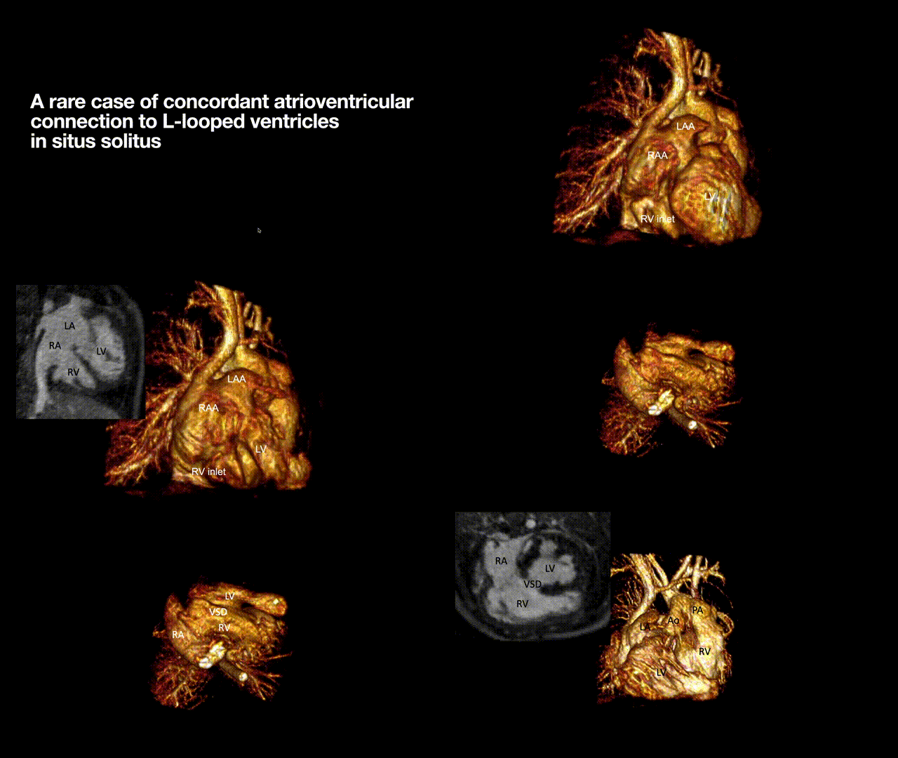
A Rare Case of Concordant Atrioventricular Connection to L-Looped Ventricles in Situs Solitus: 4-Dimensional Magnetic Resonance Imaging and 3D Printing
1
UCLA Mattel Children’s Hospital, Children’s Heart Center, Los Angeles, CA, USA
2
Diagnostic Cardiovascular Imaging Laboratory, Department of Radiology, 12222 David Geffen School of Medicine at UCLA, Los
Angeles, CA, USA
* Corresponding Author: Gregory Perens. Email:
Congenital Heart Disease 2022, 17(4), 387-392. https://doi.org/10.32604/chd.2022.021233
Received 05 February 2022; Accepted 24 May 2022; Issue published 04 July 2022
Abstract
An infant male presented with the rare anatomy consisting of situs solitus, concordant atrioventricular connections to L-looped ventricles, double outlet right ventricle (DORV), and hypoplastic aortic arch. 6 months after neonatal aortic arch repair, the morphologic right ventricle function deteriorated, and surgical evaluation was undertaken to determine if either biventricular repair with a systemic morphologic left ventricle or right ventricular exclusion was possible. After initial echocardiography, magnetic resonance imaging (MRI) was used to create detailed axial and 4-dimensional (4D) images and 3-dimensional (3D) printed models. The detailed anatomy of this rare, complex case and its use in pre-surgical planning is presented.Graphic Abstract

Keywords
Cite This Article
 Copyright © 2022 The Author(s). Published by Tech Science Press.
This work is licensed under a Creative Commons Attribution 4.0 International License , which permits unrestricted use, distribution, and reproduction in any medium, provided the original work is properly cited.


 Submit a Paper
 Propose a Special lssue
 View Full Text
 Download PDF
 Downloads
 Citation Tools