 Open Access
ARTICLE
Pulmonary Perfusion Asymmetry in Patients after Repair of Tetralogy of Fallot: A 4D Flow MRI-Based Study
1 Unité Médico-Chirurgicale de Cardiologie Congénitale et Pédiatrique, Centre de Référence des Maladies Cardiaques Congénitales Complexes, Hôpital Universitaire Necker-Enfants Malades, Université de Paris, Paris, France
2 Department of Cardiovascular and Thoracic Sciences, Fondazione Policlinico Universitario A. Gemelli IRCCS, Rome, Italy
3 Catholic University of the Sacred Heart, Rome, Italy
4 Pediatric Cardiology Department, Hospital de Santa Cruz, Centro Hospitalar Lisboa Ocidental, Lisbon, Portugal
5 Centre de Cardiologie Evecquemont, Paris, France
6 Pediatric Radiology Unit, Hôpital Universitaire Necker-Enfants Malades, Université de Paris, Paris, France
7 Decision and Bayesian Computation, Computation Biology Department, Neuroscience Department, Paris, France
8 School of Biomedical Engineering & Imaging Sciences, King’s College London, London, UK
* Corresponding Author: Francesca Raimondi. Email:
# The two authors contributed equally as first author
Congenital Heart Disease 2022, 17(2), 117-128. https://doi.org/10.32604/chd.2022.018779
Received 17 August 2021; Accepted 27 October 2021; Issue published 26 January 2022
Abstract
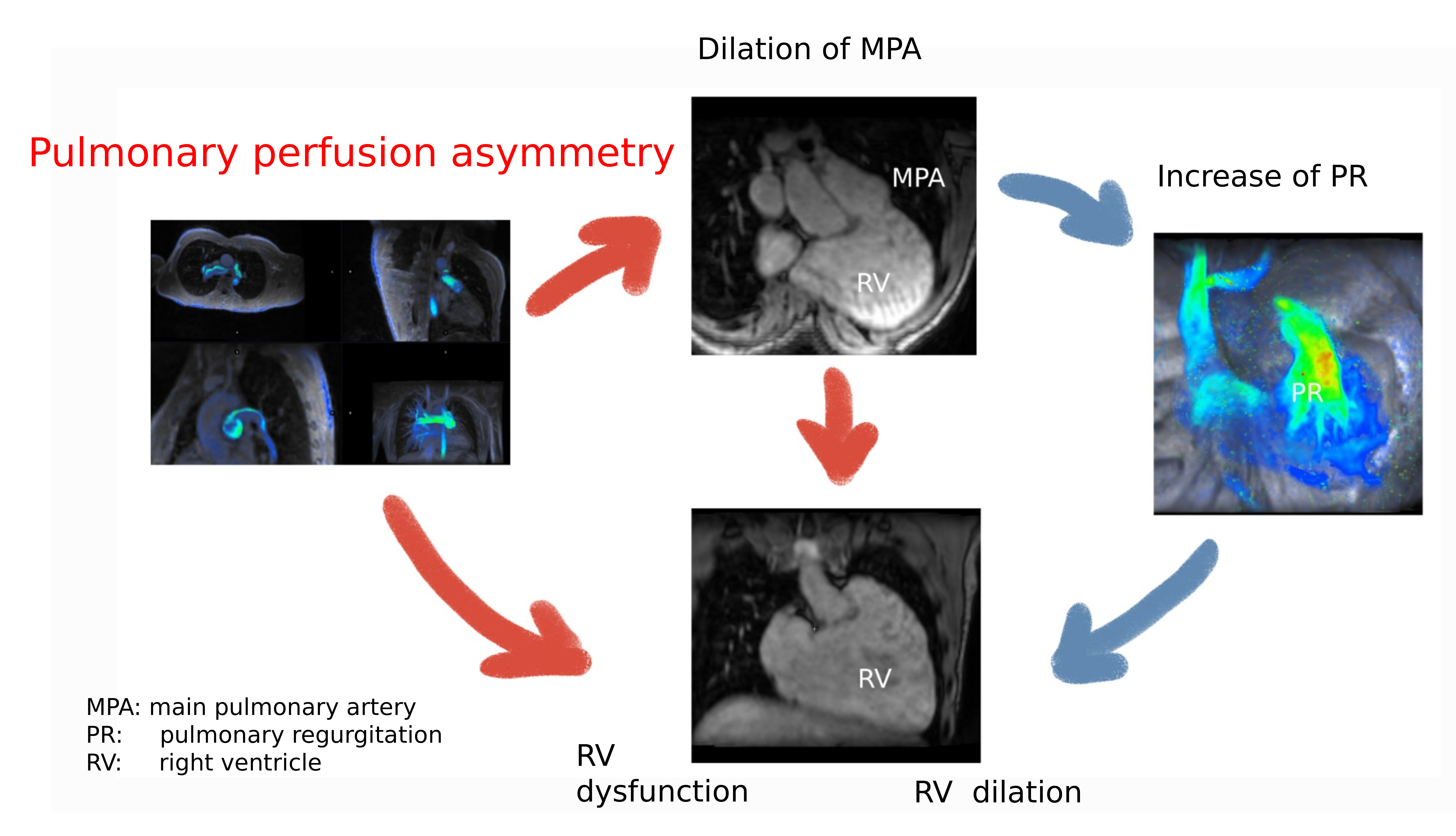
Background: Repaired Tetralogy of Fallot (rTOF) patients may have residual lesions such as main (MPA) and branch pulmonary artery stenosis (BPAS). While MPA stenosis is well studied, few data are available on BPAS in rTOF. We aimed to describe pulmonary perfusion in a large paediatric cohort of rTOF and its impact on right ventricular and outflow-tract hemodynamics using 4D flow CMR. Methods: 130 consecutive patients (mean age at CMR 14.3 ± 4.6 years) were retrospectively reviewed. 96 patients had transannular patch without valve preservation while 34 patients had conserved annulus or valved conduit. A pulmonary blood flow ratio (right pulmonary artery (RPA)/left pulmonary artery (LPA)) between 0.75 and 1.56 was considered normal. Results: Asymmetric pulmonary perfusion was present in 59/130 patients (45%), with 54/59 (91%) having left lung hypoperfusion (blood flow ratio >1.56). RPA/LPA perfusion ratio in the whole cohort was independently associated with the LPA Z-score (−0.053, p = 0.007), the RPA regurgitant fraction (RF) (0.013, p = 0.011) and previous LPA stenting (0.648, p = 0.004). Decreasing LPA % perfusion (and conversely increasing RPA % perfusion) was significantly associated with higher MPA diameter Z-score (−0.06, p = 0.007). On multivariate analysis, MPA Z-score was independently associated with pulmonary RF (0.48, p < 0.001) and with right ventricular indexed volumes (coefficient 3.6, p = 0.023). In patients with transannular patch repair, asymmetric pulmonary flow was an independent predictor of right ventricular ejection fraction (RVEF) (−3.66, p = 0.04). Conclusions: Pulmonary perfusion asymmetry is frequent in rTOF and is associated with abnormal right ventricular and outflow-tract hemodynamics, including MPA dilatation and decreased RVEF in patients after transannular patch.Graphic Abstract

Keywords
Cite This Article
 Copyright © 2022 The Author(s). Published by Tech Science Press.
This work is licensed under a Creative Commons Attribution 4.0 International License , which permits unrestricted use, distribution, and reproduction in any medium, provided the original work is properly cited.


 Submit a Paper
 Propose a Special lssue
 View Full Text
 Download PDF
 Downloads
 Citation Tools