 Open Access
CORRECTION
CORRECTION: Ameliorative effects of melatonin and zinc oxide nanoparticles treatment against adverse effects of busulfan induced infertility in male albino mice
1 Department of Zoology, Faculty of Science, Mansoura University, Mansoura, Egypt
2 The Pharmaceutical Research Institute, Albany College of Pharmacy and Health Sciences, Rensselaer, USA
3 Department of Biotechnology, Faculty of Sciences, Taif University, Taif, Saudi Arabia
* Corresponding Author: EMAN FAYAD. Email:
BIOCELL 2024, 48(12), 1835-1837. https://doi.org/10.32604/biocell.2024.054569
Issue published 30 December 2024
This article is a correction of:
Ameliorative effects of melatonin and zinc oxide nanoparticles treatment against adverse effects of busulfan induced infertility in male albino mice
Read original article
Abstract
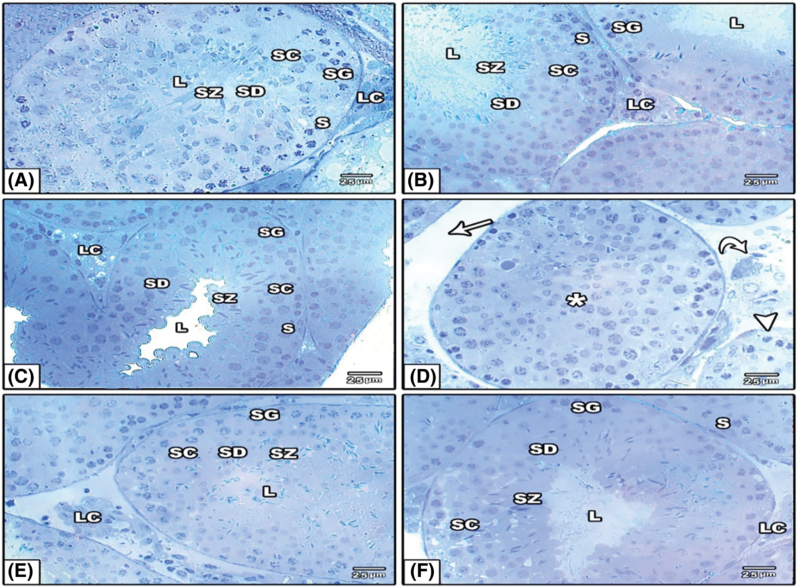
This article has no abstract.In the article “Ameliorative effects of melatonin and zinc oxide nanoparticles treatment against adverse effects of busulfan induced infertility in male albino mice” (BIOCELL, 2022, Vol. 46, No. 2, pp. 535−545. doi: 10.32604/biocell.2022.017739), an inadvertent error occurred during the compilation of Figs. 3C and 4B. This needed corrections to ensure the accuracy and integrity of the data presented.
Issue with Fig. 3C:
• Original Issue: The original compilation of Fig. 3C contained an image that was incorrectly placed due to a labeling mistake during the figure assembly process. This mislabeling did not reflect the actual experimental conditions it was meant to represent.
• Reason for Change: To accurately represent the ultrastructural changes in the testicular tissue under the specific experimental conditions (control group), we revised Fig. 3C. The new image is correctly labeled and now accurately corresponds to the correct experimental condition.
• Impact on Results: The revision of Fig. 3C does not involve any alteration of the legends or text associated with the figure. It replaces the erroneous image with the correct one, ensuring that the visual data accurately corresponds to the reported experimental findings. This correction does not impact the scientific conclusions drawn in the study.
Issue with Fig. 4B:
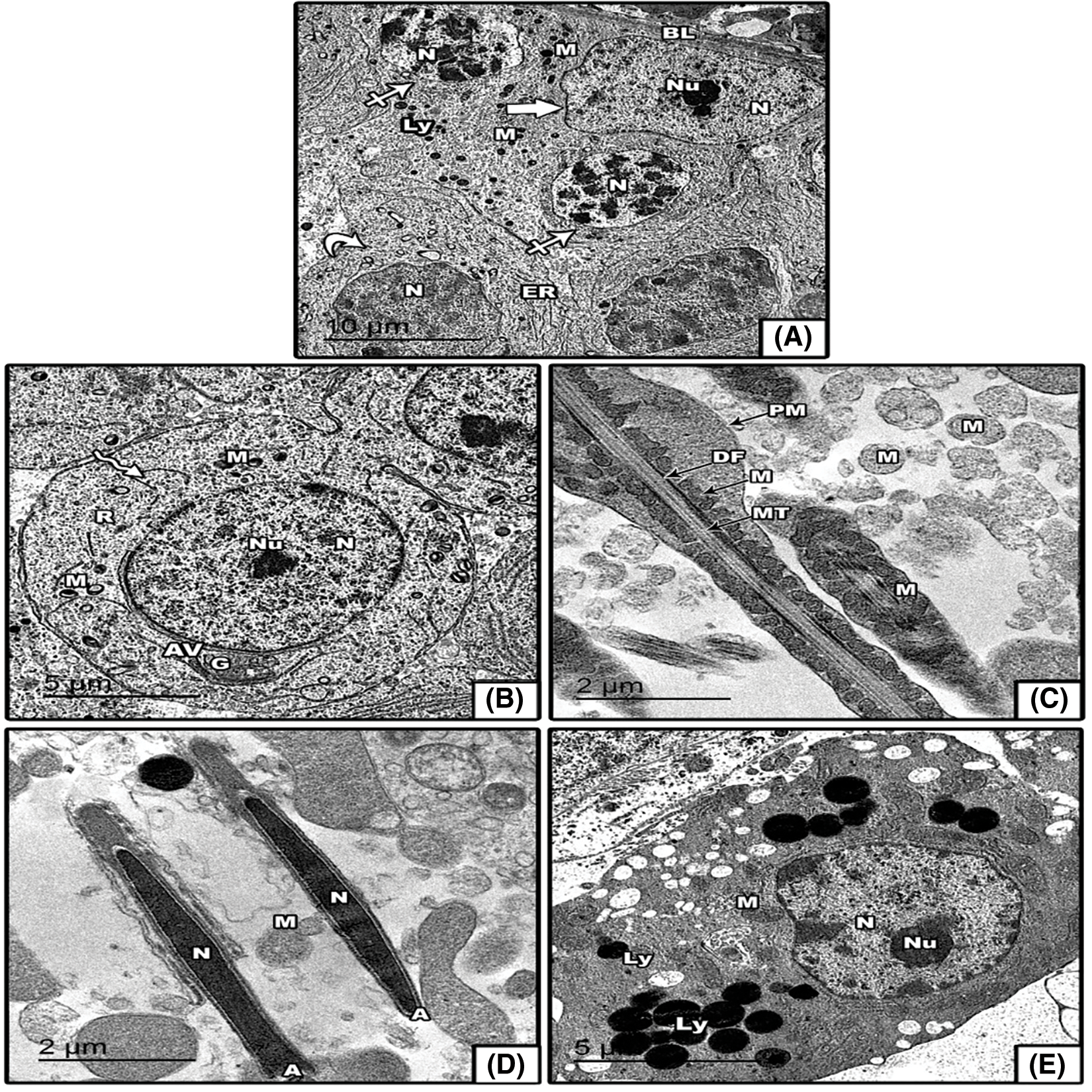
• Original Issue: The initial version of Fig. 4 contained a plate with six control images, where image B was incorrectly included. This was an oversight during the image compilation, as image B was not representative of the intended experimental condition.
• Reason for Change: To correct this error, we removed image B from the figure and adjusted the plate to include only five control images, each accurately reflecting the intended control conditions.
• Impact on Results: The removal of image B and the corresponding adjustment of Fig. 4 ensures that the Figure now accurately represents the control group. The scientific conclusions remain unchanged, as this correction was purely to align the visual data with the accurate experimental conditions.
A correction has been made in Section: Results, Sub-section: Electron microscopic observation. The corrected versions of Figs. 3 and 4 are provided. The changes were necessary to maintain the integrity of the published work and to provide accurate visual data to support the study’s findings. We confirm that these corrections do not alter any of the study’s results or conclusions, and we apologize for any inconvenience caused by the errors.
The authors would like to correct the figure as follows:


Figure 3: (A–F) Toluidine blue stained semithin sections in the testes of control and experimental groups. (A–C) control, Mel and ZnO NPs showing well organized seminiferous tubules and arrangement of Sertoli cell resting on basement membrane spermatogonia, rest on intact basement membrane, large primary spermatocytes, round and elongated spermatids and numerous mature sperms are present in the lumen, normal Leydig cells. (D) BU-treated group showing thin wall of seminiferous tubules, wide intertubular spaces with degenerated interstitial cells and reduced number of Leydig cells, no sperms found in lumen, destroyed most spermatogonia and spermatocytes cells; (E, F) Mel or ZnO NPs supplemented after BU group showing recover seminiferous tubules surrounded by regular basement membrane, improve Leydig cells, spermatogonia, Sertoli cells, and increased volume of sperms in the lumen. Abbreviations and symbols: Lumen (L), Leydig cells (LC), spermatogonia (SG), spermatozoa (SZ), separation of spermatogenic cell from the basement membrane (white arrowhead), Sertoli cells (S) and spermatocyte (SC), spermatid (SD), increased interstitial spaces (arrow), degenerated Leydig cells (curved arrows), no sperm in the lumen (*) and no spermatogenic process (arrowhead).
Electron microscopic observations: The ultrathin sections of control testis revealed that seminiferous tubules (S.T) were surrounded by thin basal lamina that contains flat myoid cells and lined by spermatogenic cells. The epithelium of spermatogenic cells showed the sequences of spermatogonia, spermatocytes, spermatids, Sertoli cells and germ cells in active spermatogenesis. Sertoli cells showed a distinct nucleus with prominent nucleoli and a cytoplasm containing numerous mitochondria, endoplasmic reticulum, lysosomes (organelles) in an active state. It is located between spermatogenic cells and rested on the basement membrane of the seminiferous tubule. The spermatogonia rested on the basal lamina of the tubule, and had a rounded nucleus with prominent nucleoli and numerous mitochondria were seen in their cytoplasm (Fig. 4A). Round spermatid appeared with regular plasma membrane surrounded by acrosomal vesicle with normal. round nucleus and nucleolus. Its cytoplasm contains well-developed mitochondria, ribosomes and Golgi apparatus (Fig. 4B). The tail of mature sperm in the middle piece showed that it is formed of central axoneme, dense outer fibers and mitochondrial sheath were around the dense fibers. The mitochondrial sheath was surrounded by the plasma membrane (Fig. 4C). The head of mature spermatozoa was elongated, containing an acrosomal cap covering the nucleus (Fig. 4D). The Leydig cell showed a regular plasma membrane with euchromatic nucleus and prominent nucleolus; its cytoplasm had many normal mitochondria and lysosomes (Fig. 4E).

Figure 4: (A–E) Electron micrographs of seminiferous tubules of control mice: (A) thin basal lamina (BL), normal appearance of Sertoli cell resting on basal lamina (Thick arrow), spermatogonia (closed arrow), Nucleus of Sertoli cell (N), nucleolus (Nu), spermatocyte (curved arrow), lysosomes (Ly), mitochondria (M), endoplasmic reticulum (ER), (B) Spermatid (zigzag arrow), Nucleus of spermatid (N), nucleolus (Nu), Acrosomal vesicle (AV), mitochondria (M), Ribosome (R), Golgi apparatus (G); (C) Plasma membrane (PM), Dense fiber (DF), Microtubule (MT), mitochondria (M); (D) Acrosome (A), Nucleus of sperm (N); (E) Nucleus of Leydig cell (N) with nucleolus (Nu), Lysosomes (Ly), mitochondria (M).
Cite This Article
 Copyright © 2024 The Author(s). Published by Tech Science Press.
This work is licensed under a Creative Commons Attribution 4.0 International License , which permits unrestricted use, distribution, and reproduction in any medium, provided the original work is properly cited.


 Submit a Paper
 Propose a Special lssue
 View Full Text
 Download PDF
 Downloads
 Citation Tools